Follow us on Google News (click on ☆)

In a resting state, meaning in the absence of external stimuli or tasks performed by the individual, certain groups of neurons exhibit synchronous activity resulting from connections with other brain areas. Mapping these different connections constitutes functional networks known as "resting-state" and forms the functional connectome.
These networks, which are involved in functions such as memory consolidation or processing visual or auditory data, evolve throughout life and can be altered by neurological diseases. These factors should therefore be considered for the correct interpretation of functional MRI (fMRI) images, especially when establishing treatment for patients with neurological diseases or who have suffered a stroke.
A team from the Center for Biological and Medical Magnetic Resonance (CRMBM, CNRS/Aix-Marseille University), in collaboration with the Institute of Systems Neuroscience (INS, Aix-Marseille University/Inserm), conducted a pilot study revealing the impact of sex, age, and the day/night cycle on the resting-state brain connectivity of mice observed by fMRI.
The fMRI, sensitive to local variations in blood flow and oxygenation induced by neuron activation, highlights the networks of neurons activated simultaneously in the resting state. Adult male and female mice, aged 5 and 14 months, were examined in resting-state fMRI at four moments of the day/night cycle (10:00 AM, 4:00 PM, 9:00 PM, 4:00 AM).

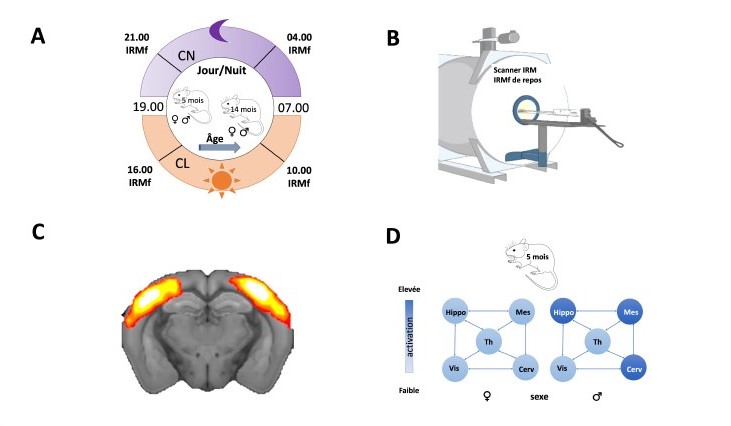
(A-B) fMRI study of the effects of the day/night cycle, sex, and age on the resting-state functional connectome of the mouse. Mice were studied at 2 different ages (5 and 14 months) and at 4 moments of the day/night cycle (10:00 AM, 4:00 PM, 9:00 PM, 4:00 AM) on an MRI scanner.
(C) The sensorimotor network involved in sensory perception and motor activity is one of the known functional networks identified in CRMBM's study.
(D) Example showing the effect of sex on one of the new networks identified by CRMBM: increased brain activity in memory-related structures in males compared to females at 5 months old, regardless of lighting conditions. CL = light condition, CN = night condition, Hippo = hippocampus, Mes = midbrain, Th = thalamus, Vis = visual cortex, Cerv = cerebellum.
© Armelle Lokossou, CRMBM/INS.
The results revealed 16 resting-state functional networks, including 5 new ones, and demonstrate the influence of the studied variables on the functional connectome of mice. Thus, fMRI images showed that the activity of a network involved in memorization was stronger in male mice than in female mice (all 5 months old), regardless of light conditions.
Furthermore, in 14-month-old female mice (the experiment could not be conducted with males), a significant increase in pineal gland activity was observed during periods of darkness. The pineal gland controls the production of melatonin, a hormone that plays a central role in regulating biological rhythms.
This initial study demonstrates the necessity to consider parameters such as age, sex, and the day/night cycle when interpreting resting-state fMRI images in mice used in research as models to study alterations in the functional connectome in diseases such as epilepsy or Alzheimer's disease.
References:
Impact of the day/night cycle on functional connectome in ageing male and female mice.
Houéfa Armelle Lokossou, Giovanni Rabuffo, Monique Bernard, Christophe Bernard, Angèle Viola, Teodora-Adriana Perles-Barbacaru.
NeuroImage, 15 April 2024, 120576.
https://doi.org/10.1016/j.neuroimage.2024.120576
Article available on the open archive platform HAL