Follow us on Google News (click on ☆)

Pixabay illustration image
A team from the University of Geneva (UNIGE) has identified a mechanism by which thalamic projections target specific neurons and alter their excitability. This work, published in Nature Communications, reveals a novel way of communicating between two regions of the brain, the thalamus and the somatosensory cortex. It could explain why the same sensory stimulus does not always cause the same sensation and open avenues for better understanding certain mental disorders.
The same sensory stimulus can sometimes be perceived clearly or remain blurry. This phenomenon is explained by the way the brain integrates these perceptions. Thus, touching an object outside our field of vision may be enough to identify it, or not. These perceptual variations remain poorly understood, but they could depend on factors such as attention or the disruptive presence of other stimuli.
What is certain, according to neuroscientists, is that when we touch something, the sensory signals from the skin's receptors are interpreted by a specialized region of the brain called the "somatosensory cortex."
This alteration could play a role in certain pathologies, such as autism spectrum disorders.
Along the way, these signals pass through a complex network of neurons, including a crucial brain structure called the "thalamus," which acts as a relay. However, this process is not one-way. A significant part of the thalamus also receives feedback signals from the cortex and redistributes them, forming a feedback loop. But the exact role and functioning of this loop remain poorly understood.

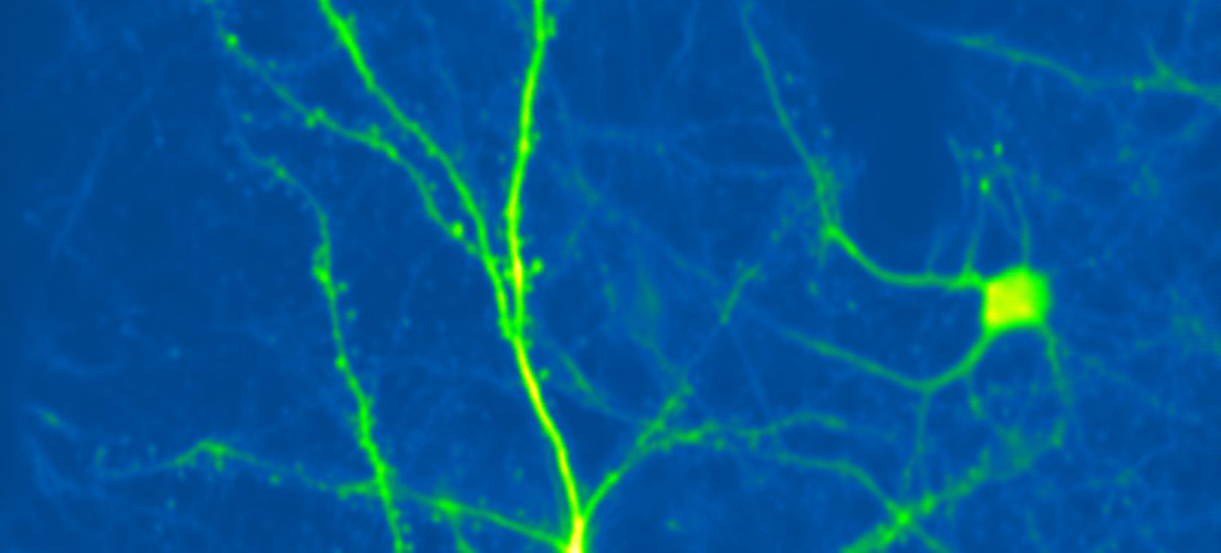
Cortical neuron expressing a green fluorescent protein, imaged in the living mouse brain using two-photon microscopy.
© Ronan Chéreau
A new modulatory pathway
To explore this question, the UNIGE neuroscientists studied a region located at the top of pyramidal neurons in the somatosensory cortex, rich in dendrites - extensions of neurons that receive electrical signals from other neurons.
"Pyramidal neurons have somewhat surprising shapes. They are asymmetrical, both in their shape and their functions. What happens at the top of the neuron is different from what happens at the bottom," explains Anthony Holtmaat, Full Professor in the Department of Fundamental Neurosciences and at the Synapsy Center for Research in Mental Health Neurosciences at the UNIGE Faculty of Medicine, who led the study.
His team focused on mice and a particular neural pathway, in which the top of pyramidal neurons receives projections from a specific part of the thalamus. By stimulating the animal's whiskers - the equivalent of touch in humans - a precise dialogue between these projections and the dendrites of the pyramidal neurons could be highlighted.
"What is remarkable, unlike the thalamic projections known until now for activating pyramidal neurons, is that this part of the thalamus, operating in a feedback loop, modulates their activity, notably by making them more sensitive to stimuli," says Ronan Chéreau, Senior Assistant in the Department of Fundamental Neurosciences and co-author of the study.
An unexpected receptor
Thanks to cutting-edge techniques - imaging, optogenetics, pharmacology, and especially electrophysiology - the team was able to record the electrical activity of tiny structures like dendrites. These different approaches made it possible to understand how this modulation occurs at the synaptic level.
Normally, the neurotransmitter glutamate acts as an activation signal. It helps neurons transmit sensory information by triggering an electrical response in the next neuron. But in this new mechanism, the glutamate released by the thalamic projections acts differently. It binds to another type of receptor, located in a specific region of the cortical pyramidal neuron.
In response, the latter changes its reactivity state rather than being directly excited. The neuron thus becomes more easily activated, as if it were being primed to better respond to a future sensory stimulus.
"This is a previously unknown pathway for modulation. Usually, the modulation of pyramidal neurons is ensured by the balance between excitatory and inhibitory neurons, not by this type of mechanism," specifies Ronan Chéreau.
Many implications
By showing that a specific feedback loop between the somatosensory cortex and the thalamus can modulate the excitability of cortical neurons, the study suggests that thalamic pathways do not merely transmit sensory signals, but also act as selective amplifiers of cortical activity.
"In other words, our perception of touch is not only shaped by incoming sensory data, but also by the dynamic interactions within the thalamocortical network," adds Anthony Holtmaat.
Furthermore, this mechanism could contribute to understanding the perceptual flexibility observed in sleep or wakefulness states, during which sensory thresholds vary. Its alteration could also play a role in certain pathologies, such as autism spectrum disorders.