Follow us on Google News (click on ☆)

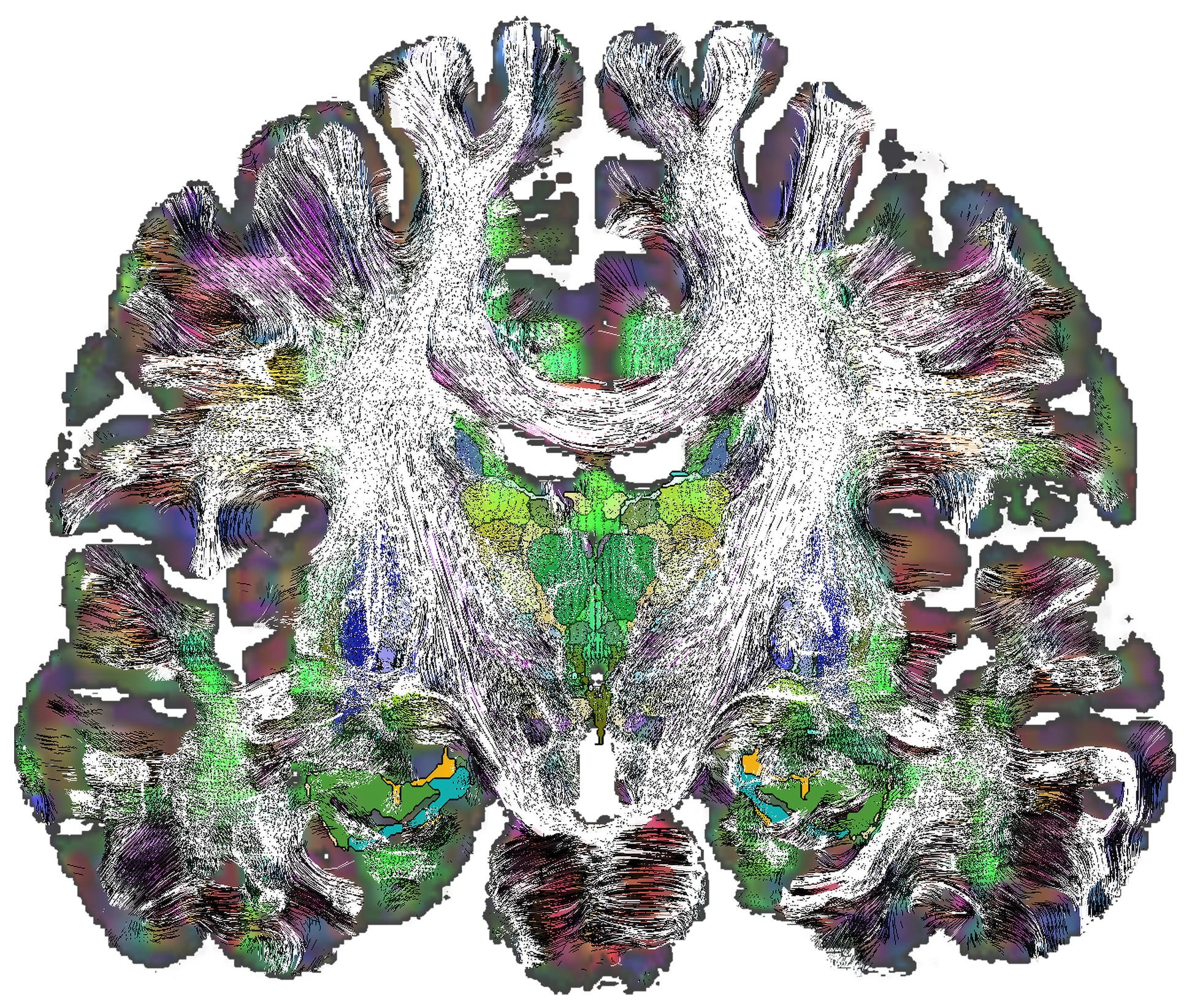
Coronal section (vertical, anterior view) passing through the middle of the deep brain. It combines, over a 1 mm thickness: in the background, an orientation map of nerve fibers with color coding (blue: vertical; red: right-left/left-right; green: anteroposterior/posteroanterior), a representation of deep structures (black outlines and colored surfaces), and tracings of nerve fiber bundles from diffusion imaging (fine white lines) revealing the complexity of the connectome.
© J.J. Lemaire/Institut Pascal
Understanding the deep structures of the brain is essential for comprehending its function and pathologies, as well as for identifying new therapeutic targets and developing brain stimulation treatments for neurodegenerative diseases, particularly Parkinson's disease. This architecture remains poorly understood, partly due to the complexity of its 3D organization and the terminology used to describe the structures, which is often incomplete and imprecise.
In a transdisciplinary approach, a team led by the Institut Pascal (IP, CNRS/Université Clermont-Auvergne), involving neurosurgeons and specialists in imaging and digital sciences, has produced an advanced mapping of the human deep brain. This new deep brain atlas is made available to researchers across all disciplines (neuroscience, biology, computer science, clinical practice, etc.).
To precisely map the structures of the deep brain and their interconnections at a submillimeter scale, the team conducted a set of MRI scans on a healthy volunteer. Multiple imaging modalities (multimodal MRI) were used to highlight fine structures (by enhancing image contrast) as well as the architecture of connections between different cellular nuclei (by observing the diffusion of water molecules in tissues).
The integration of these different acquisitions resulted in a detailed atlas of 118 deep brain structures and their connections, accompanied by comprehensive terminology (location, orientation, etc.) to enable future users to practically utilize this new research tool.
These results were validated by comparison with a broad set of data, including histological section images.
Since the study's publication, the deep brain atlas, available as open-source, has been downloaded over a thousand times by researchers from various disciplines. The new tool can still be improved by increasing the resolution of the mapping to observe finer architectural details. Additionally, a software environment will be developed to facilitate its use by researchers and clinicians.
This new tool is made available to researchers in neuroscience, biology, computer science, and clinical practice. It could aid in the development of treatments for neurological diseases.