Detection of bioluminescence deep within the brain
Follow us on Google News (click on ☆)

Scientists often label cells with luminescent proteins to track their development or observe changes in genetic expression. However, these techniques have so far been limited in exploring deep brain structures, as light scatters too much before becoming detectable.
Engineers at MIT, led by Alan Jasanoff, have innovated by making blood vessels in the brain capable of detecting this light. By expressing a bacterial protein, these vessels enlarge in the presence of light, a change observable by MRI.
This method, called bioluminescence imaging using hemodynamics or BLUsH, could allow the study of changes in genetic expression, the anatomical connections between cells, or how cells communicate with each other.

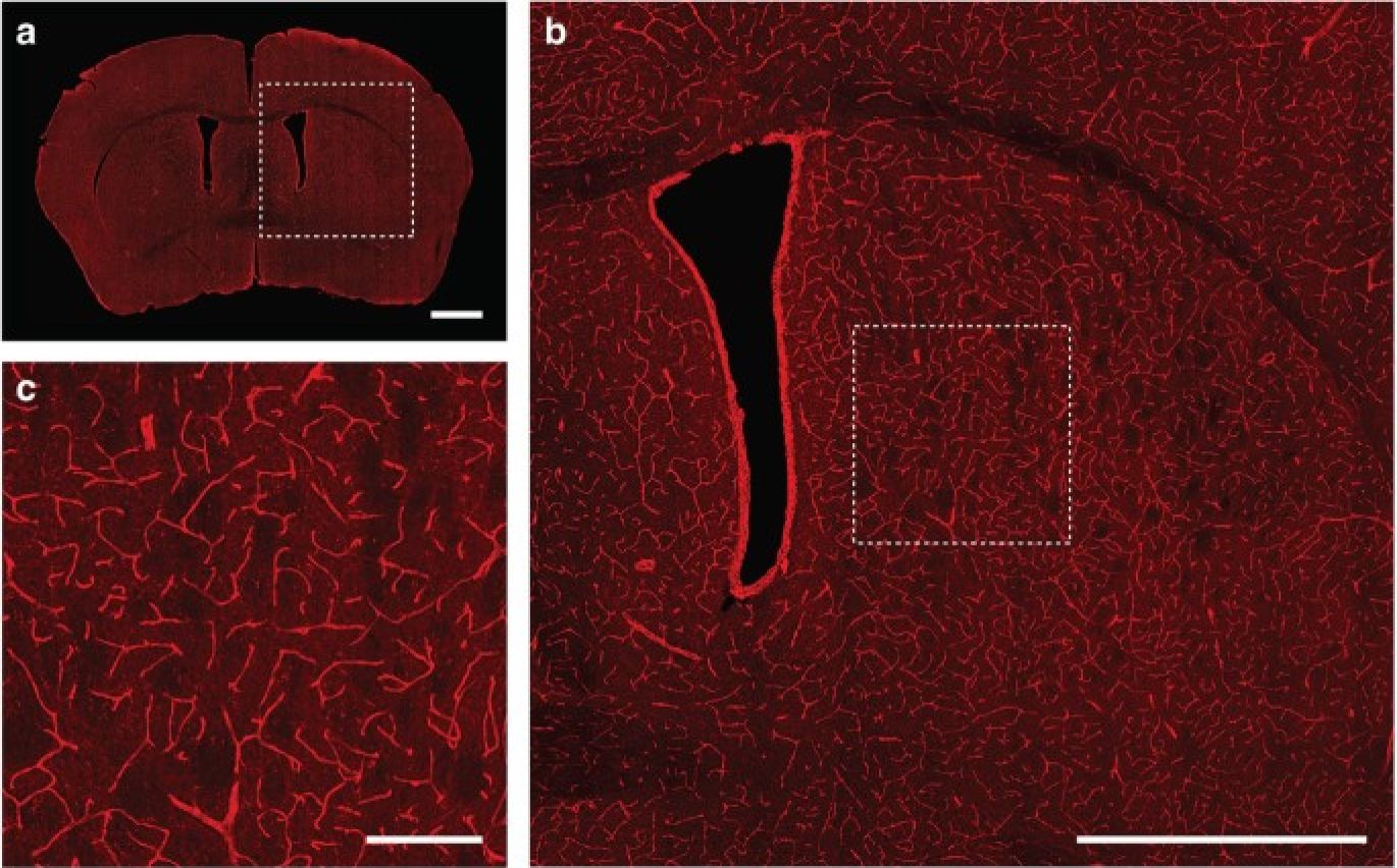
A new method for detecting bioluminescence in the brain uses magnetic resonance imaging (MRI). Developed at MIT, this technique could allow researchers to explore the brain's internal mechanisms in more detail than ever before. Blood vessels appear bright red after transduction with a gene that confers photosensitivity.
The study, published in Nature Biomedical Engineering, opens new perspectives for brain research, particularly in rodents, with the hope of extending the application to other animal models.