Follow us on Google News (click on ☆)
Traditional obstacles in organ imaging could soon disappear. Thanks to an approach based on ionic liquids, scientists can now make tissues transparent while keeping their structure intact.

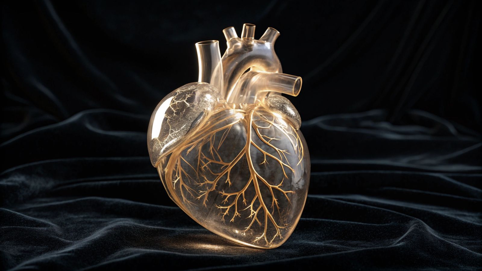
Fictional illustration image
The principle of ionic transparency
The ionic liquids used possess a unique property: they penetrate tissues without deforming them. Unlike classical methods, they avoid problems of expansion or crystallization during cooling.
This technique, named VIVIT, transforms biological samples into a glassy state. Organs treated this way become translucent, allowing precise microscopic observation. Fluorescent markers also gain in intensity, revealing previously invisible details.
Applications are immediate for neural mapping. Initial experiments on human and murine brains have already yielded promising results. The method could extend to other organs, such as the heart or spleen.
Notable practical advantages
Unlike existing techniques, VIVIT fully preserves tissue morphology. Treated samples withstand time, storing without damage under extreme cold conditions.
The fluorescence enhancement facilitates the identification of cellular connections. Rare proteins or neural networks become easily observable. This unmatched precision particularly interests research in personalized medicine.
Scientists are already considering clinical applications. The detection of pathological markers or artificial intelligence-assisted imaging could benefit from this innovation.